
Which Organ Sits In The V Part Of The Ribs / Rib Cage Rib Cage Bones
Skeletal System Ribs Ribs The ribs partially enclose and protect the chest cavity, where many vital organs (including the heart and the lungs) are located. The rib cage is collectively.
Human Anatomy Ribs Pictures How Many Ribs do Humans Have? Bodytomy
The rib cage or thoracic cage (called thorax) is located in the most medial portion of the body and protects vital organs like the heart and lungs. The rib cage is an integral part of the axial skeleton in vertebrates and it lies in the thoracic cavity.

Rib Cage Muscles / Thoracic Muscles Attachments Actions Teachmeanatomy
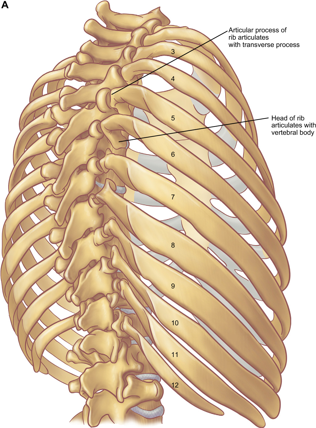
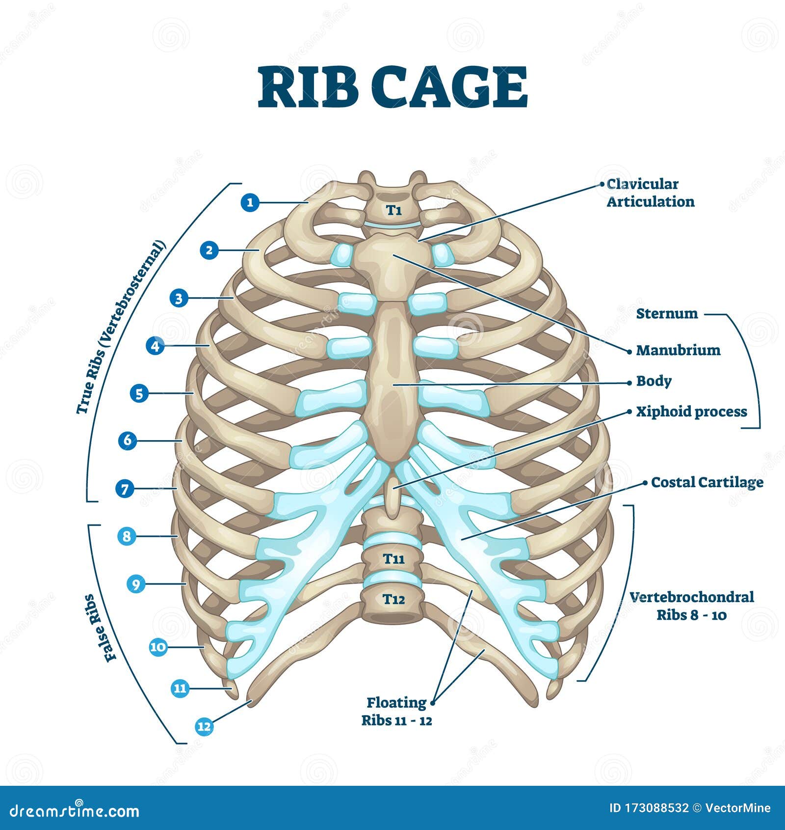
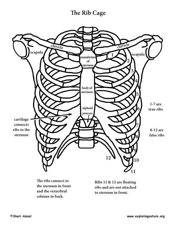
Introduction to the Structure of the Ribcage and Ribs: There are twelve (12) pairs of ribs and all articulate posteriorly with the thoracic vertebrae. Lateral view of a pair of ribs articulating with the thoracic vertebrae. The true ribs ( ribs 1-7) attach to the sternum by costal cartilages.

Diagram Rib Cage With Organs / Anatomy Under Ribs Human Body Anatomy
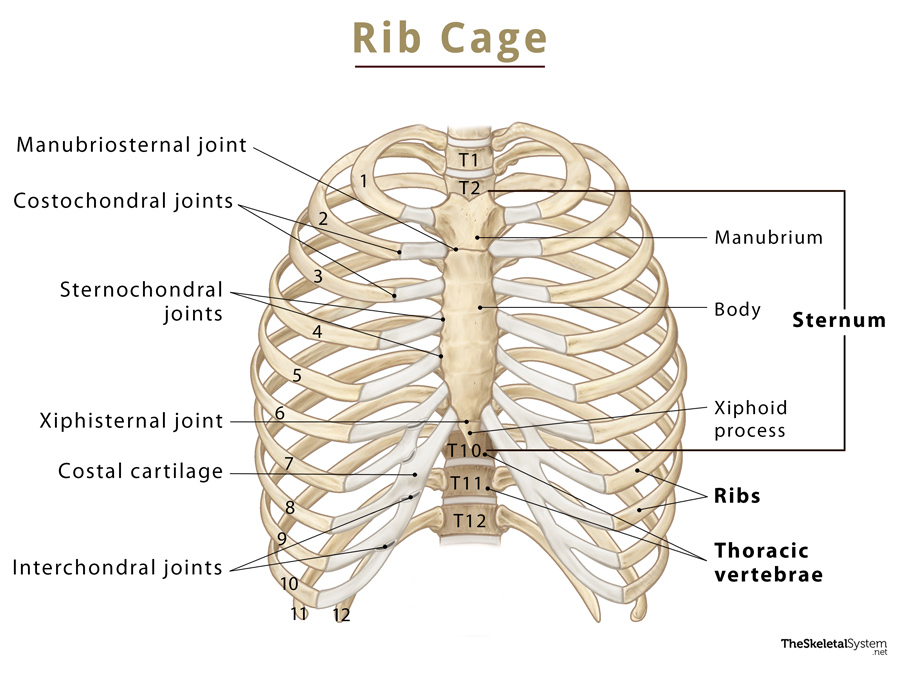
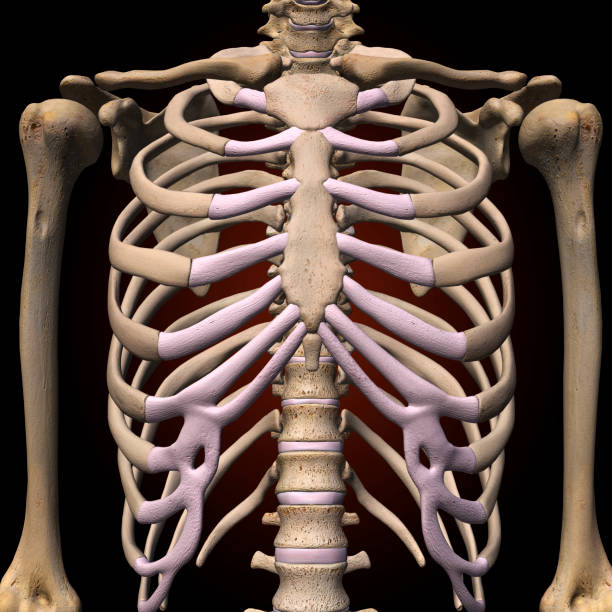
The thoracic cage is a bony case consisting of ribs and sternum which encases vital organs like the lungs and the heart and shapes the chest. There are 12 pairs of ribs in the body. They are attached to the vertebral column with most of the ribs anteriorly joining to the sternum bone via costal cartilage. The first seven ribs directly attach to.

Rib Cage Medical Art Library
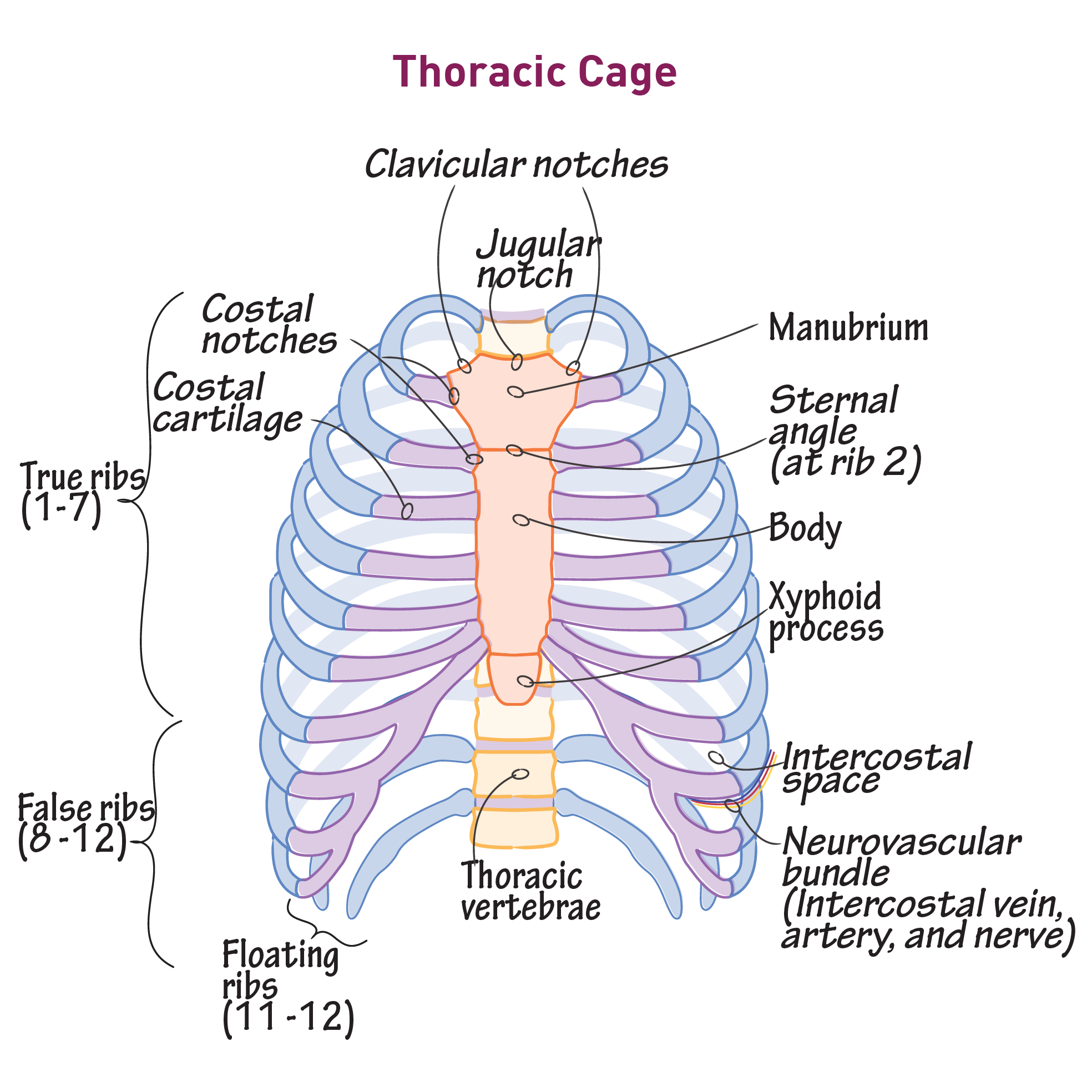
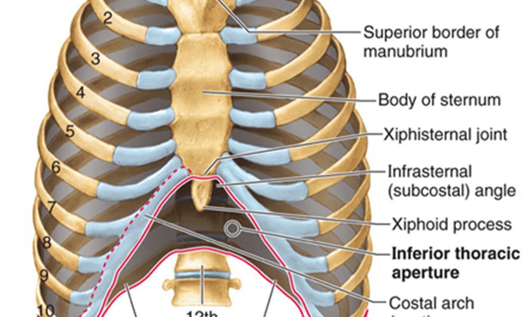
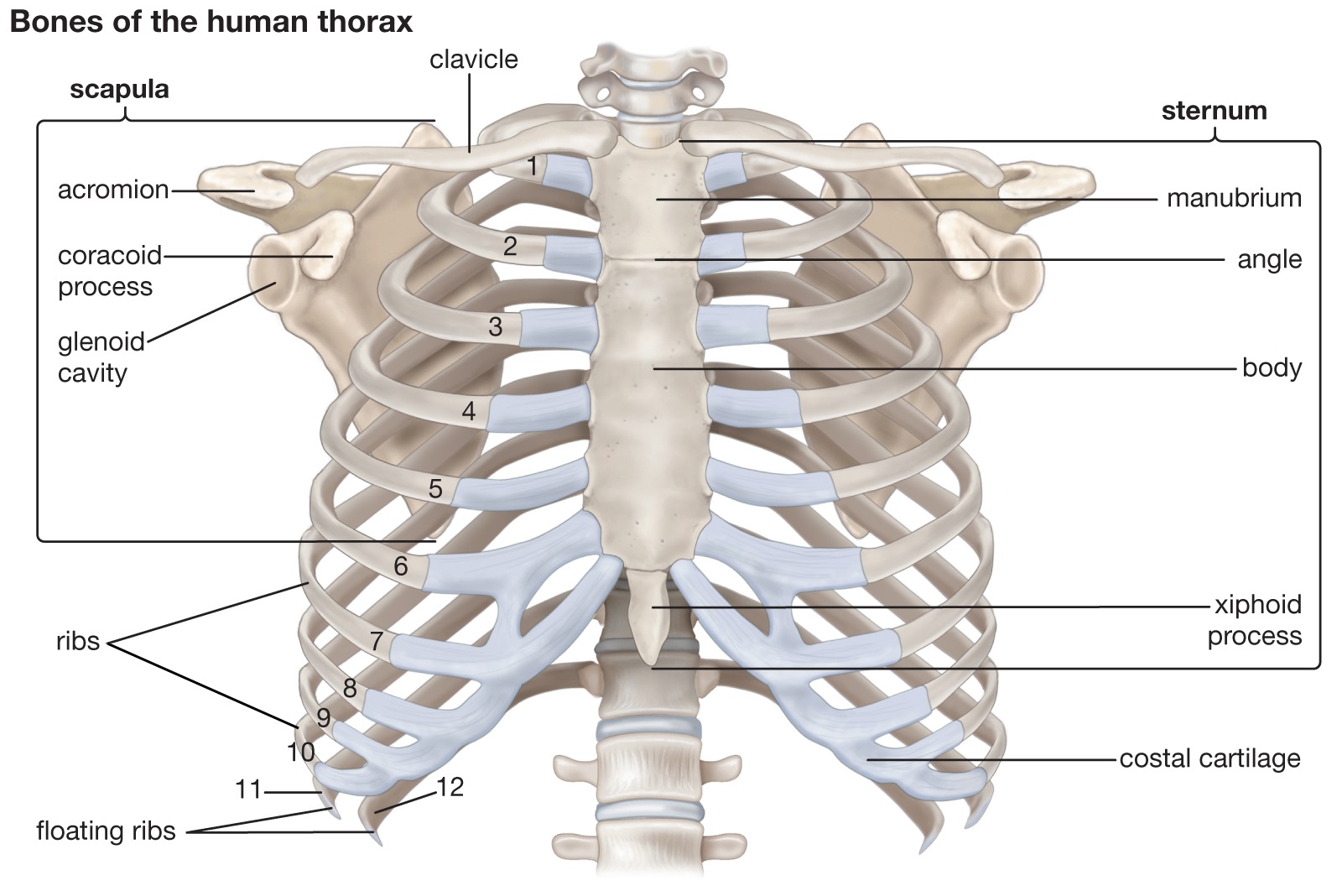
The thoracic cage protects the heart and lungs. Figure 7.32 Thoracic Cage The thoracic cage is formed by the (a) sternum and (b) 12 pairs of ribs with their costal cartilages. The ribs are anchored posteriorly to the 12 thoracic vertebrae. The sternum consists of the manubrium, body, and xiphoid process. The ribs are classified as true ribs (1.
Rib Cage Anatomy Posterior Medical Animation from Visual Health
thoracic vertebra torso rib cage, in vertebrate anatomy, basketlike skeletal structure that forms the chest, or thorax, and is made up of the ribs and their corresponding attachments to the sternum (breastbone) and the vertebral column.

Human Rib Cage Drawing at GetDrawings Free download
The rib cage is part of the axial skeleton. The average human is born with the same number of ribs regardless of gender. The ribs articulate with the thoracic vertebra. For example, the first rib.
Rib Cage Names of Bones, Anatomy, Functions, & Labeled Diagram
The diaphragm and intercostal muscles control these movements. They also provide points of origin or attachment for several essential muscles in the chest and thoracic wall. Rib bones participate in erythropoiesis (the process of producing red blood cells). They are one of the sites where erythropoiesis occurs throughout your adult life. Anatomy

Rib cage diagrams atilaassist
In this video we discuss the structure of the rib cage or thoracic cage. We cover the different bones that make up the rib cage and some of the functions of.
Find Out 50+ Truths On Anatomy Diagram Rib Area Your Friends Missed to
The thoracic cage (rib cage) forms the thorax (chest) portion of the body. It consists of the 12 pairs of ribs with their costal cartilages and the sternum (Figure 6.5.1 6.5. 1 ). The ribs are anchored posteriorly to the 12 thoracic vertebrae (T1-T12). The thoracic cage protects the heart and lungs.
Rib Cage Anatomy, Labeled Vector Illustration Diagram Stock Vector
The main bones in the abdominal region are the ribs. The rib cage protects vital internal organs. There are 12 pairs of ribs and they attach to the spine. There are seven upper ribs, known as.
Back Rib Cage Muscles Rib Cage Muscles Pin On Back Pain As in the
The ribs are curved, flat bones which form the majority of the thoracic cage. They are extremely light, but highly resilient; contributing to their role in protecting the internal thoracic organs.
Anatomy Of Rib Cage Rib Cage Diagram With Organs Human Anatomy Body
Numbering order of the vertebrae of the human spinal column There are thirty-three vertebrae in the human vertebral column. The rib cage is associated with TH1−TH12. Ribs are described based on their location and connection with the sternum. All ribs are attached posteriorly to the thoracic vertebrae and are numbered accordingly one to twelve.
Rib cage diagram Healthiack
Shoulder & Back Arm & Hand Pelvis & Hip Leg & Foot has three important functions: protection, support and respiration. The bones of the rib cage are the thoracic vertebrae and the 12 pairs of is a flat bone that is made up of three parts, the (1) manubrium,, and the ( 3) xiphoid process.

diagram of the ribs
The thoracic cage (rib cage) forms the thorax (chest) portion of the body. It consists of the 12 pairs of ribs with their costal cartilages and the sternum ( Figure 7.5.1 ). The ribs are anchored posteriorly to the 12 thoracic vertebrae (T1-T12). The thoracic cage protects the heart and lungs.
Rib Cage Yoga and Medical Science
The rib cage, also known as the thoracic cage, is the bony structure that shapes and protects the thoracic cavity and the organs in it. It has a conical shape and looks somewhat like a birdcage (hence the name). This part of the axial skeleton is located in the chest, above the abdominal cavity. R i b C a g e Functions